Blogger templates

Pages

Showing posts with label Anatomy; Joints. Show all posts

Thursday, September 2, 2010

Joints of the Upper Limb

Shoulder Joint
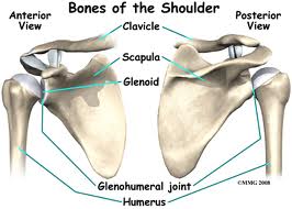
                         The synovial  ball and socket variety of joint is formed by the glenoid cavity of the scapula and head of the humerus. It is the most mobile joint of the body.This joint is also known as glenohumeral joint. The joint has the following characteristics.
  • Articular Capsule 
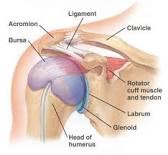
                        The joint surface is covered with the sleeve of hyaline cartilage called as articular capsule. The articular capsule starts from the superior border of glenoid cavity and ends at the anatomical neck of the humerus. The inferior portion of the capsule is very weak.
  • Glenohumeral Ligament
                         This ligament starts from the glenoid cavity and ends at the anatomical neck of the humerus. It is made up of three bands of fibrous tissue.
  • Coracohumeral ligament  
                         It starts from the coracoid process of humerus and ends at the greater tuberosity of the humerus. It strengthens the shoulder joint.
  • Transverse Humeral Ligament
                         It starts from the greater tuberosity and ends at the lesser tuberosity. This ligament provide stability to the shoulder joint.
  • Glenoid Lambrum
                         The glenoid rim is the fibro-cartilage which deepens the shoulder joint and also provide extra stability to the shoulder joint.
  • Bursa
                         There are four bursa present around the shoulder joint.
  1.    Sub-acromial bursa
  2.    Sub-coracoid bursa
  3.    Sub-scapular bursa
  4.    Sub-deltoid bursa
  • Movements
              Flexion:
                            Anterior fibres of deltoid
                            Pectoralis major
                            Biceps
                            Coracobrachialis
              Extension:
                                Posterior fibres of deltoid
                                 Latissimus dorsi
                                Teres major
              Abduction:                             
                                Middle fibres of deltoid
                                Supraspinatus
              Adduction:
                                Pectoralis major
                                 Latissimus dorsi
                                Teres major
                                Teres minor
              Lateral rotation:
                                 Infraspinatus
                                 Teres minor
              Medial rotation:
                                 Subscapularis
                                 Latissimus dorsi
                                 Teres major


Elbow Joint
                         The synovial hinge joint formed by the articulation of trochlear notch of ulna and head of radius with trochlea and capitulum of the humerus. It is extremely stable joint of the body having the following characteristics.
  • Articular Capsule
                         The joint surface is covered by the articular capsule which starts anterioly from the radial and coronoid fossa and ends at the annular ligament and coronoid process. The articular capsule protects the inner parts of the elbow joint.
  • Annular Ligament
                          The annular ligament is round in shape which surrounds the radial head.
  • Ulnar Collateral Ligament
                          It starts from the medial epicondyle and ends at the coronoid and olecranon processes of ulna.
  • Radial Collateral Ligament
                          It starts from the lateral epicondyle and ends at the annular ligament and radial notch of ulna.
  • Movements
             Flexion:
                           Brachialis
                           Biceps brachii
                           Brachioradialis
                           Pronator teres    
             Extension:
                           Triceps
                            Anconeus

Proximal Radioulnar Joint
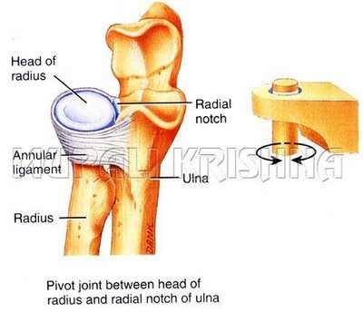
                       The  pivot type of synovial joint formed by the articulation of radial head with radial notch of ulna.
Distal Radioulnar Joint
                       The pivot type of  synovial joint which is formed by the articulation of ulnar head with ulnar notch of radius.
  • Movements
             Pronation:
                               Pronator teres
                                Pronator quadratus
             Supination:
                                Biceps brachii
                                 Supinator  


Wrist Joint
                      The condyloid type of synovial joint formed by the distal end of the radius and the articular disc of the distal radioulnar joint with the proximal row of carpal bones except the pisiform i.e scaphoid, lunate and triquetrium.
  • Movements
              Flexion/extension  
              Radial (abduction) deviation
              Ulnar (adduction) deviation
              Circumduction 
    
Carpometacarpal and Intermetacarpal Joints
                      These all are plane synovial joints except that of thumb which is saddle joint.
  • Movements
               Only Gliding

Metacarpophalangeal Joints
                       These are condyloid type of synovial joints which allow movement in two directions.
  • Movements 
               Flexion
               Extension
               Abduction
               Adduction
               Circumduction

Interphalangeal Joints
                           These are uniaxial hinge joints which allow movement in only on direction.
  • Movements
              Flexion
             Extension


Pectoral Girdle
                            The pectoral girdle(clavicle and scapula) connects the upper limb to the trunk, therefore, its articulations are includeed with those of upper limb.
Sternoclavicular Joint
                             This is a saddle type of synovial joint and is the only bony articulation between the upper limb and the axial skeleton.The medial end of the clavicle articulates with the manubrium of the sternum.
  • Movements
             Allows movements of the pectoral girdle and upper limb. In flexion of the upper limb, the SC joint (i.e., clavicle on sternum) moves anteriorly, posteriorly and inferiorly.

Acromioclavicular Joint
                                This is a plane type of synovial joint. Acromial end of the clavicle articulates with the acromion of the scapula.
  • Movements
              The acromion of the scapula rotates on the acromial end of the clavicle.

Sunday, August 22, 2010

Joints of the Lower Limb

Hip Joint
          It is synovial ball and socket variety of joint. It is formed by the articulation of head of femur with the acetabulum of hip bone. The following are the characteristics of hip joint.

  • Articular Capsule
          Superiorly it attaches to the acetabular rim(edge) and then encircle the hip joint in the form of circular fibers upto the neck of the femur.
  • Ileofemoral Ligament
          It is consist of longitudinal fibres which start from the anterior inferior iliac spine and extend upto the intertrochanteric line anteriorly and intertrochanteric crest posteriorly.
  • Pubofemoral Ligament
          It is also consist of logitudinal fibers which start from the pubic rim and converge on the intertrochanteric line anteriorly.
  • Ischiofemoral Ligament 
          Its longitudinal fibers starts from the ischium posteriorly and converge on the intertrochanteric crest of the femur.
  • Ligament of head of Femur (Ligamentus teres or Round ligament)
          It starts from the acetabular fossa superiorly and attaches to the fovea capitus inferiorly.
  • Acetabular Labrum
          The cavity of the acetabulum contains the soft cartilaginous structure called labrum which deepens the acetabular cavity thus adds the stability of hip joint.
  • Transverse Ligament
          It is consist of circular fibers which starts from acetabuar notch and attaches to the ligamentus teres and to the neclk of femur.

  • Movements
          • Flexion
             o Iliopsoas
             o Rectus femoris
             o Sartorius

           • Extension
              o Gluteus maximus
              o Hamstring muscles

           • Abduction
              o Gluteus medius 
              o Gluteus minimus

           • Adduction
              o Adductor longus
              o Adductor fibres of the adductor magnus
              o Adductor brevis

           • Lateral rotation
              o Piriformis
              o Obturator internus and externus
              o Quadratus femoris
              o Superior and inferior Gamelli

           • Medial rotation
              o Anterior fibres if gluteus medius
              o Gluteus minimus
              o Tensor fasciae lata

Knee Joint
          It is the most largest and complex joint of the body. The knee is made up of four main bones- the femur (thigh bone), the tibia (shin bone), fibula (outer shin bone) and patella (kneecap). It is made up of three compartments.
                                              
Medial Compartment: It includes medial condyles of tibia and femur along with medial meniscus.
Lateral Compartment: It includes lateral condyles of femur and tibia along with lateral meniscus.
Anterior Compartment: It includes intercondylar area of femur and posterior surface of patella.

Extra-articular Features:
  • Articular capsule
          It does'nt cover knee joint completely. It is combination of different ligaments, tendons and their fibers.
  • Medial and Lateral Patellar Retinaculae
          The tendons of the quadricep femoris form retinaculae on both sides of the patella giving strength to the knee joint.
  • Patellar Ligament
          It connects the base of patella to tibial tuberosity. It is also giving strength to the knee joint anteriorly. Infra patellar pad of fat separates the patellar ligament from the cavity of knee joint.

  • Medial Collateral Ligament
           Superiorly starts from the medial condyle of femur and attaches to the medial condyle of tibia.It give strength to knee joint from medial side.
  • Lateral Collateral Ligament
          Superiorly starts from the lateral condyle of femur and attaches to the head of fibula.
  • Oblique Popliteal Ligament 
          It is flat ligament which extends from the intercondylar fossa of femur to the head of tibia.This ligament covers knee joint from the back.
  • Arcuate Popliteal Ligament
          It starts from the lateral condyle of femur and extends to the styloid process of fibula. This ligament covers the lower back of knee joint.

Intra-articular Features:
  • Cruciate Ligaments
          They are named due to cross shaped appearance. Anterior and posterior cruciate ligaments are named due to their relation with intercondylar area of the tibia.

               Anterior Cruciate Ligament (ACL): It starts from the anterior of intercondylar area of tibia, proceeds laterally and attaches to the medial surface of lateral condyle of femur. the point of attachment is b/w lateral meniscus and posterior cruciate ligament. It restiicts hyperextention of knee joint and also prevents anterior sliding of tibia over femur.

                Posterior Cruciate Ligament (PCL): It starts posterior to the intercondylar area of tibia, proceeds medially and attaches to the lateral surface of the medial condyle of femur. It prevents posterior sliding of tibia over femur.
  • Menisci (Sing- Meniscus)
          There are two fibrocartilages discs present on the upper surface of tibial plateau.
               Medial Meniscus: It is C-shaped disc. Its anterior part lies infront of ACL while posterior part part attaches infront of Lateral meniscus . Medial meniscus is attach medially to the medial collateral ligament and it also attaches the ACL.

               Lateral Meniscus: it is almost complete circular shape. It is starts posterior to the ACL and posteriorly attaches infront of medial meniscus.

               Function of Menisci: It make superior surface of the tibia concurrent to the articular surface of femur and also help in the distribution of synovial fluid in the knee joint cavity.
  • Movements
           • Extension
             o Quadriceps Femoris

           • Flexion

              o Hamstring muscles


Ankle Joint
          The synovial hinge joint.It is formed by the articulation of lower end of tibia, the two malleoli and body of talus.
  • Articular Capsule
          Capsule is thin and allows large range of movement.
  • Ligaments
          Capsule is strengthened by ligaments

               o Medial or deltoid ligament

               o Anterior talofibular ligament

               o Calcaneofibular ligament

               o Posterior talofibular ligament

  • Movements
      • Dorsiflexion
         o Tibialis anterior
         o Extensor hallucis longus
         o Extensor digitorum longus
         o Peroneus tertius

      • Plantar flexion
         o Gastrocnemius
         o Soleus
         o Plantaris
         o Peroneus longus
         o Peroneus brevis
         o Tibialis posterior
         o Flexor digitorum longus
         o Flexor hallucis longus

Inversion and eversion take place at the tarsal joints.

Subtalar Joint
          The joint between talus and calcaneum is called as subtalar joint. Gliding and rotatory movements are possible.


Design by Free WordPress Themes | Bloggerized by Lasantha - Premium Blogger Themes | Facebook Themes