Blogger templates

Pages

Showing posts with label Anatomy; Muscles. Show all posts

Thursday, September 2, 2010

Skull and Facial Muscles

  • Frontalis muscle
Origin:
          Epicranial aponeurosis

Insertion:
          Supra orbital margin

Function:
          Raises the upper eyelids, and wrinkles the skin of the forehead

  • Occipitalis muscle
Origin:
          Posterior margin of the occipital bone and mastoid process of temporal bone

Insertion:
          Epicranial aponeurosis

Function:
          Draws skin of the scalp backward

Facial Muscles

  • Orbicularis oris muscle
Origin:
          Upper and lower margins of lips

Insertion:
          Angle of the mouth

Function:
          Closes the mouth

  • Orbicularis oculi muscle
Origin:
          Upper and lower margins of the orbit

Insertion:
          Angle of the orbits

Function:
          Closes the eye

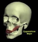
  • Zygomatic major muscle
Origin:
          Zygomatic bone

Insertion:
          Angle of the mouth

Function:
          Raises the upper lip

  • Zygomatic minor muscle
Origin:
          Zygomatic bone

Insertion:
          Lower lip

Function:
          Depression of lower lip

  • Levator labii superioris muscle
Origin:
          Upper lip

Insertion:
          Angle of the mouth

Function:
          Raises the upper lip

  • Depressor labii inferioris muscle
Origin:
          Lower lip

Insertion:
          Angle of the mouth

Function:
          Depression of lower lip

  • Levator palpebrii superioris muscle
Origin:
          Roof of the orbit and lesser wing of the sphenoid bone

Insertion:
          Skin of the superior eyelid

Function:
          Elevates the upper eyelid

  • Buccinator muscle
Origin:
          Alveolar processes of maxillae and mandible

Insertion:
          Upper and lower lips

Function:
          Approximation of upper and lower lips as in ‘whistling’

  • Risorius muscle
Origin:
          Fascia over the parotid gland

Insertion:
          Lower lip

Function:
            Draw the mouth backward

  • Mentalis muscle
Origin:
          Angle of mandible

Insertion:
          Mental foramen

Function:
          Movement of mandible

  • Corrigurator supercilli muscle
Origin:
          Medial side of supra orbital margin

Insertion:
          Eyebrow

Function:
          Movement of eyebrow

Saturday, August 28, 2010

Muscles of the Lower Limb

Muscles of Gluteal region


  • Gluteus Maximus Muscle
          This is the largest and superficial muscle of the body mainly responsible for the prominence of Gluteal region.

Origin:
          Outer surface of ileum behind the posterior Gluteal line, coccyx and sacrum

Insertion:
          Ileotibial tract and gluteal tuberosity

Innervation:
          Inferior Gluteal nerve

Function:
          Extension and laterally rotation of thigh at hip joint and also extension of the knee joint through ileotibial tract

  • Gluteus Minimus Muscle
Origin:
          Outer surface of ileum b/w middle Gluteal line and inferior Gluteal line

Insertion:
          Anterior border of greater tronchanter of femur

Innervation:
          Superior gluteal nerve

Function:
          Abducts and medially rotates the thigh at hip joint


  • Gluteus Medius Muscle
Origin:
          Outer surface of ileum between iliac crest above and middle gluteal line below

Insertion:
          Lateral surface of greater tronchanter

Innervation:
          Superior gluteal nerve

Function:
          Abducts the thigh and anterior fibers medially rotate the thigh at hip joint

  • Tensor Fasciae Latae Muscle
Origin:
          Iliac crest b/w ASIS and iliac tubercle

Insertion:
          Ileotibial tract

Innervation:
          Superior gluteal nerve

Function:
           Assists gluteus maximus in extending the knee through ileotibial tract and also abducts the thigh along with medius and minimus muscles

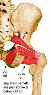
  • Piriformis Muscle
Origin:
          Anterior surface of the 2nd, 3rd and 4th sacral vertebrae in pelvis

Insertion:
          Upper border of greater tronchanter of femur

Innervation:
          1st and 2nd sacral nerves

Function:
          Lateral rotation of the thigh at hip joint

  • Obturator Internus Muscle
Origin:
          Inner surface of Obturator membrane

Insertion:
          Upper border of greater tronchanter
Innervation:
          Obturator internus from sacral plexus

Function:
          Lateral rotation of the thigh at hip joint

  • Gamellus Superior Muscle
Origin:
          Spine of ischium

Insertion:
          Upper border of greater tronchanter of femur

Innervation:
          Obturator internus from sacral plexus

Function:
          Lateral rotation of thigh at hip joint

  • Gamellus Inferior Muscle
Origin:
          Upper margin of ischial tuberosity

Insertion:
          Upper border of greater tronchanter of femur

Innervation:
          Sacral plexus

Function:
          Lateral rotation of thigh at hip joint

  • Quadratus Femoris Muscle
Origin:
          Lateral border of ischial tuberosity

Insertion:
          Quadrate tubercle of femur

Innervation:
          Sacral plexus

Function:
          Lateral rotation of thigh at hip joint

Muscle of Anterior compartment of Thigh


  • Sartorius Muscle
Origin:
           ASIS (Anterior Superior Iliac Spine)

Insertion:
          Upper medial border of tibia

Innervation:
          Femoral nerve

Function:
          Flexes, abducts and laterally rotates the thigh at hip joint. It flexes and medially rotates the leg at the knee joint.

  • Iliacus Muscle
Origin:
          Iliac fossa within abdomen

Insertion:
          Lesser tronchanter of femur

Innervation:
          Femoral nerve

Function:
         Flexes the thigh on abdomen and abdomen on thigh as sitting up from lying

  • Psoas Muscle
Origin:
          Transverse processes and intervertebral discs of T12 to L5

Insertion:
          Lesser tronchanter of femur

Innervation:
          Lumber plexus

Function:
          Flexion of thigh on abdomen and abdomen on thigh as in sitting up from lying


  • Pectineus Muscle
Origin:
          Superior ramus of pubic bone

Insertion:
          Upper border of linea aspera

Innervation:
          Femoral nerve

Function:
          Flexion and adduction of thigh at hip joint

  • Quadriceps Femoris Muscle
          It is consist of the following four parts having same quadriceps tendon which inserts on patella forming patellar ligament. Patellar ligament in turn inserts on the tibial tuberosity.


Rectus Femoris

Origin:
   Straight head:
          ASIS

   Reflected head:
          Ileum above acetabulum

Insertion:
          Inserts on patella as quadriceps tendon which in turn inserts on tibial tuberosity

Innervation:
          Femoral nerve

Function:
          Flexion of thigh at hip joint and extension of hip at knee joint


Vastus Medialis


Origin:
          Upper medial shaft of femur

Insertion:
          Quadriceps tendon
Innervation:
          Femoral nerve

Function:
          Extension of leg at knee joint


Vastus Lateralis


Origin:
          Upper lateral shaft of femur
Insertion:
          Quadriceps tendon

Innervation:
          Femoral nerve

Function:
          Extension of leg at knee joint


Vastus Medius


Origin:
          Anterior and lateral shaft of femur

Insertion:
          Quadriceps tendon

Innervat:ion:
          Femoral nerve

Function:
          Extension of leg at knee joint


Muscles of Posterior compartment of Thigh
          These muscles are also called as Hamstring muscles
Biceps Femoris Muscle
Origin:
   Long head:
          Ischial tuberosity

   Short head:
          Linea aspera

Insertion:
          Head of fibula

Innervation:
          Sciatic nerve

Function:
          Extension of thigh at hip joint and flexion along with lateral rotation of leg at knee joint


Semitendinosus Muscle

Origin:
          Ischial tuberosity
 Insertion:
            Upper medial surface of shaft of tibia


Innervation:
   Tibial portion of sciatic nerve
Function:
          Extension of thigh at hip joint; Flexion and medial rotation of leg at knee joint

Semimembranosus Muscle

Origin:
          Ischial tuberosity

Insertion:
          Medial condyle of tibia

Innervation:
          Tibial portion of sciatic nerve

Function:
          Extension of thigh at hip joint; flexion and medial rotation of leg at knee joint


Adductor Magnus (hamstring portion)
Origin:
          Ischial tuberosity

Insertion:
          Adductor tubercle of femur

Innervation:
          Sciatic nerve

Function:
          Extension of thigh at hip joint


Muscles of Medial compartment of Thigh

  • Gracilis Muscle

Origin:

          Inferior ramus of pubis and ramus of ischium

Insertion:

          Upper medial border of tibia

Innervation:

          Obturator nerve

Function:

          Adduction of thigh at hip joint and flexion of leg at knee joint


  • Adductor Brevis Muscle

Origin:

          Inferior ramus of pubis

Insertion:

          Linea aspera

Innervation:

          Obturator nerve

Function:

          Adduction of thigh at hip joint

  • Adductor Longus Muscle

Origin:

          Body and inferior ramus of pubis

Insertion:

          Linea aspera

Innervation:

          Obturator nerve

Function:

          Adduction of thigh at hip joint


  • Adductor Magnus Muscle
Origin:
          Inferior ramus of pubis, ramus of ischium and ischial tuberosity

Insertion:
   Adductor part:
          Linea aspera

   Hamstring part:
          Adductor tubercle

Innervation:
   Adductor portion:
          Obturator nerve

   Hamstring portion:
          Sciatic nerve

Function:
   Adductor portion:
          Adduction of thigh at hip joint

   Hamstring portion:
          Extension of leg at knee joint


  • Obturator Externus Muscle


Origin:
          Outer surface of Obturator membrane

Insertion:
          Medial border of greater tronchanter

Innervation:
          Obturator nerve

Function:
          Lateral rotation of thigh at hip joint


Muscles of Anterior compartment of Leg

  • Tibialis Anterior Muscle
Origin:
          Shaft of tibia and interosseous membrane

Insertion:
          Medial cuneiform and base of 1st metatarsal

Innervation:
          Deep peroneal nerve

Function:
          Extension of foot at ankle joint; inversion of foot at subtalar and transverse tarsal joints; holds up medial longitudinal arch of the foot

  • Extensor Digitorum Longus Muscle
Origin:
          Anterior surface of shaft of fibula
Insertion:
          Extensor expansion of lateral four toes

Innervation:
          Deep peroneal nerve

Function:
          Extension of toes and extension of foot at ankle joint

  • Peroneus Tertius Muscle
Origin:
          Anterior surface of shaft of fibula

Insertion:
          Base of distal phalanx of fifth toe

Innervation:
          Deep peroneal nerve
Function:
          Extension of foot at ankle joint; eversion of foot at subtalar joint and extension of 5th toe

  • Extensor Hallucis Longus Muscle
Origin:
          Anterior surface of shaft of fibula

Insertion:
          Distal phalanx of big toe

Innervation:
          Deep peroneal nerve

Function:
          Extension of foot at ankle joint and extension o big toe

  • Extensor Digitorum Brevis Muscle
Origin:
          Calcaneum

Insertion:
          Proximal phalanx of great toe, 2nd, 3rd and 4th toes

Innervation:
          Deep peroneal nerve

Function:
          Extension of toes except the 5th

Muscles of Lateral compartment of Leg

  • Peroneus Longus Muscle
Origin:
          Lateral surface of shaft of fibula

Insertion:
          Medial cuneiform and base of 1st metatarsal

Innervation:
          Superficial peroneal nerve

Function:
          Plantar flexion of ankle joint and also support the medial longitudinal arch of foot

  • Peroneus Brevis Muscle
Origin:
          Lateral surface of shaft fibula

Insertion:
          Base of 5th metatarsal bone

Innervation:
          Superficial peroneal nerve

Function:
          Plantar flexion of ankle joint and longitudinal arch of the foot

Muscles of Posterior compartment of Leg

Superficial Group

  • Gastrocnemius Muscle
Origin:
   Lateral head:
          Lateral condyle of femur

   Medial head:
          Medial condyle of femur

Insertion:
          Achilles tendon which in turn inserts on the posterior surface of calcaneum

Innervation:
          Tibial portion of sciatic nerve

Function:
          Flexion of the knee joint and plantar flexion of the ankle joint

  • Plantaris Muscle
Origin:
          Just above the supracondylar ridge of femur
Insertion:
          Achilles tendon

Innervation:
          Tibial nerve (branch of sciatic nerve)

Function:
          Flexion of leg at knee joint and plantar flexion of foot at ankle joint

  • Soleus Muscle
Origin:
          Shaft of tibia (soleal line) and fibula
Insertion:
          Achilles tendon

Innervation:
          Tibial nerve

Function:
          Plantar flexion of ankle joint


Deep Group

  • Popliteus Muscle
Origin:
          Lateral surface of lateral condyle of femur

Insertion:
          Just above soleal line on posterior surface of tibia

Innervation:
          Tibial nerve

Function:
          Flexion and medial rotation of leg at knee joint

  • Flexor Hallucis Longus Muscle
Origin:


          Posterior surface of fibula

Insertion:
          Base of distal phalanx of big toe

Innervation:
          Tibial nerve

Function:
          Flexion of big toe and flexion of ankle joint

  • Flexor Digitorum Longus Muscle
Origin:
          Posterior surface of shaft of tibia
Insertion:
          Distal phalanges of lateral four toes

Innervation:
          Tibial nerve

Function:
          Flexion of lateral four toes and flexion of ankle joint

  • Tibialis Posterior Muscle
Origin:
          Posterior surface of shaft of tibia fibula and interosseous membrane

Insertion:
          Navicular and other adjoining bones (neighboring)

Innervation:
          Tibial nerve

Function:
          Plantar flexion of ankle joint, support the transverse arch and inverts foot at subtalar and transverse tarsal joint


Monday, August 16, 2010

Muscles of the Upper Limb

Muscles connecting Upper Limb to the Thorax
  • Pectoralis Major Muscle
            Origin: It originates from the medial third of the clavicle, sternum and upper six costal cartilages.
             Insertion: It inserts at the lateral lip of bicipital groove of the humerus.

             Function: Abduction and medial rotation of the arm.
             Nerve Supply: Medial and Lateral pectoral Nerves from Brachial Plexus

  • Pectoralis Minor Muscle
            Origin: It originates from the 3rd to 5th ribs.

             Insertion: It inserts at the coracoid process of the scapula.

             Function: Depression of the scapula.

             Nerve Supply: Medial Pectoral Nerve from Brachial Plexus

  • Subclavius Muscle
             Origin: It originates from the superior border of the 1st rib.

             Insertion: It inserts at the clavicle.

             Function: Depression of the clavicle.

             Nerve Supply: Nerve to subclavius from Upper Trunk of Brachial Plexus
  • Serratus Anterior Muscle
             Origin: It originates from the 1st eight ribs.

             Insertion: It inserts at the medial border of the scapula.

             Function: Upward, downward and medial rotation of the scapula.

             Nerve Supply: Long Thoracic Nerve


Muscles connecting Scapula to the Vertebral Column

  • Trapezius Muscle
             Origin: Ocipital bone, spines of 7th cervical  and all thoracic vertebrae.

             Insertion: Lateral third of clavicle, spine of scapula and acromion process.

             Function: Elevation, depression rotation of the scapula.

             Nerve Supply: Accessory nerve and C3 and C4

  • Latissimus Dorsi Muscle
              Origin: Iliac crest, Spines of lower six thoracic vertebrae, lower three or four ribs and inferior angle of scapula.
              Insertion: Floor of bicipital groove of Humerus.

              Function: Extention, Adduction and Medial rotation of the arm.

              Nerve Supply: Thoracodorsal Nerve

  • Levator Scapulae Muscle
              Origin: It originates from the transverse processes of upper three cervical vertebrae.

              Insertion: It inserts at the medial upper border of the scapula.

              Function: It elevates the scapula.

              Nerve Supply: C3,C4 and Dorsal Scapular Nerve

  • Rhomboid Minor Muscle
              Origin: It originates from the spinous processes  of the 7th and T1 vertebrae.

              Insertion: It inserts on the superior medial border of the scapula.

              Function: Elevation and medial rotation of the scapula.

              Nerve Supply: Dorsal Scapular Nerve
  • Rhomboid Major Muscle 
              Origin: It originates from the spinous processes of 2nd to 5th thoracic vertebrae.

              Insertion: It inserts on the medial lower border of the scapula.

              Function: Elevation and medial rotation of the scapula.

              Nerve Supply: Dorsal Scapular Nerve


Muscles connecting Scapula to the Humerus
  • Supra spinatus Muscle
             Origin: It originates from the supra spinatus fossa of the scapula.

             Insertion: It inserts on the greater tubercle of the humerus.
          
             Function: Abduction and stabilization of the shoulder joint.

             Nerve Supply: Suprascapular nerve
  • Infra spinatus Muscle
              Origin: It originates from the infraspinatus fossa of the scapula.
              Insertion: It inserts on the greater tuberosity of the humerus.
              Function: Lateral rotation of the arm and stabilization of the shoulder joint.

              Nerve Supply: Suprascapular nerve

  • Sub scapularis Muscle
              Origin: It originates from the sub scapular fossa of the scapula.

              Insertion: It inserts on the lesser tuberosity of the humerus.

              Function: Medial rotation of the arm and staabilization of the shoulder joint.

              Nerve Supply: Upper and Lower subscapular Nerves

  • Teres Minor Muscle
               Origin: It originates from the inferior lateral border of the scapula.

               Insertion: It inserts on the greater tuberosity of the humerus.
               Function: Lateral rotation of the arm.

               Nerve Supply: Axillary Nerve
  • Teres Major Muscle
               Origin: It originates from the inferior lateral border of the scapula.

               Insertion: It inserts on the medial lip of bicipital groove.

               Function: Medial rotation of the arm and stabilization of the shoulder joint.

               Nerve Supply: Lower Subscapular Nerve


  • Deltoid Muscle
             Origin: It originates from the lateral third of the clavicle, acromion process and spine of the scapula.

             Insertion: It inserts on the deltoid tuberosity of the humerus.

             Function: The anterior fibers flex, middle fibers abduct and posterior fibers extend the shoulder joint.

             Nerve Supply: Axillary Nerve

Muscles of the Arm 


  • Coracobrachialis Muscle
              Origin: It originates from the coracoid process of the scapula.

              Insertion: It inserts on the medial third of the humerus.

              Function: Flexion of the arm.

              Nerve Supply: Musculocutaneous Nerve

  • Brachialis Muscle
              Origin: It originates from the lower 1/3 of the anterior aspect of humerus.

              Insertion: It inserts on the coronoid processs of the ulna.

              Function: Flexion of the elbow joint and supination of the forearm.

              Nerve Supply: Musculocutaneous Nerve


  • Bicep Brachii Muscle
              Origin: The long head of bicep muscle originates from the supra glenoid tubercle of the glenoid cavity while short head from the corocoid process of the scapula.

              Insertion: Radial tuberosity

              Function: Flexion of the elbow joint and supination of the forearm.

              Nerve Supply: Musculocutaneous Nerve


  • Tricep Brachii Muscle
              Origin: Long head originates from the infraglenoid tubercle of the glenoid cavity, medial head from medial side below the radial groove and lateral head from
 lateral side above the radial groove.

              Insertion: Olecranon process of the ulna.

              Function: Extention of the elbow joint.

              Nerve Supply: Radial Nerve


Muscles of the Forearm

A. Anterior Surface of Forearm (flexors primarily)

  • 1. Superficial Group
a. Pronator teres:

b. Flexor Carpi Radialis: flexes wrist and abducts hand

c. Palmaris Longus:

d. Flexor Carpi Ulnaris: flexes wrist and adducts hand

  • 2. Intermediate Group
a. Flexor Digitorum Superficialis: flexes wrist and flexes digits 2 - 5 at the MP and PIP joints
(MP=metacarpal-phalangeal PIP=proximal interphalangeal)

  • 3. Deep Group
a. Flexor Digitorum Profundus: flexes wrist and digits 2-5 at the MP, PIP and DIP joints (DIP=distal interphalangeal)

b. Flexor Pollicis Longus:

c. Pronator quadratus:

Innervation of Anterior compartment muscles: All the muscles are suplied by the Median nerve and its branches except the flexor carpi ulnaris and the medial part of the flexor digitorum profundus, which are supplied by the Ulnar nerve.

B. Lateral Surface of Forearm

a. Extensor Carpi Radialis Longus: extends wrist and abducts hand

b. Brachioradialis:

Nerve Supply to Lateral compartment muscles: Radial Nerve


C. Posterior Surface of Forearm (extensors primarily)

  • 1. Superficial Group
a. Anconeus:

b. Extensor Carpi Radialis Brevis

c. Extensor Digitorum: extends wrist and extends digits 2-5 at MP, PIP, and DIP joints

d. Extensor Carpi Ulnaris:

e. Extensor digiti minimi:

  • 2. Deep Group
a. Supinator: supinates forearm

b. Abductor Pollicis Longus:

c. Extensor Pollicis Longus: extends thumb

d. Extensor Pollicis brevis:

e. Extensor indicis:

Innervation to Posterior compartment muscles: Deep branch of radial nerve.


Selected Muscles
  • Brachioradialis Muscle
              Origin: Lateral supracondylar ridge of humerus.

              Insertion: Base of styloid process of radius.

              Function: Flexion of the forearm at elbow joint.

              Innervation: Radial Nerve

  • Anconeus Muscle

             Origin: Lateral epicondyle of humerus.

             Insertion: Lateral surface of olecranon process of ulna.

             Function: Extention of elbow joint.

             Nerve Supply: Deep branch of Radial Nerve


  • Supinator Muscle

              Origin: Lateral epicondyle of humerus.

              Insertion: Neck and shaft of radius.

              Function: Supination of the forearm.

             Nerve Supply: Deep branch of Radial Nerve


  • Pronator Teres Muscle
              Origin: Humeral head originates from the medial epicondyle of humerus and ulnar head from the medial border of coronoid process of ulna.

              Insertion: Lateral aspect of shaft of radius.

              Function: Pronation and flexion of the forearm.

              Nerve Supply: Median Nerve

  • Pronator Quadratus Muscle
              Origin: Anterior surface of shaft of ulna.

              Insertion: Anterior surface of shaft of radius.

              Function: Pronation of forearm.

              Nerve Supply: Median Nerve


 HAND MUSCLES


A. Thenar muscles: move the thumb:
                                                                             Short muscles of thumb innervated by Median Nerve except adductor pollicis, which is by deep branch of Ulnar Nerve.


B. Hypothenar muscles:
                                        Short Muscles of Little Finger are innervated by deep branch of Ulnar Nerve.


C. Midpalmar muscles

1. All of these muscles flex the MP joints and extend the PIP/DIP joints


a. Lumbricales: (4)
                          First two (lateral) are innervated by Median Nerve and third and fourth by deep branch of Ulnar Nerve.


b. Palmar Interossei: (4) :
                                    Innervated by deep branch of Ulnar Nerve


c. Dorsal Interossei: (4) :
                                   Innervated by deep branch of Ulnar Nerve


Design by Free WordPress Themes | Bloggerized by Lasantha - Premium Blogger Themes | Facebook Themes